Review Article | | Peer-Reviewed

Feline Herpesvirus: A Persistently Relevant Disease, from Infection to Clinical Management

Received: 20 February 2025     Accepted: 3 March 2025     Published: 15 September 2025
Views:       Downloads:
Abstract

Feline herpesvirus type 1 (FHV-1) is a widespread pathogen responsible for feline viral rhinotracheitis, a highly contagious disease affecting cats worldwide. This virus primarily targets the upper respiratory tract and ocular tissues, leading to clinical signs such as sneezing, nasal discharge, conjunctivitis, keratitis, and, in severe cases, corneal ulceration. FHV-1 is particularly problematic in young, immunocompromised, or stressed cats, and latent infections can result in recurrent clinical manifestations. The pathogenesis of FHV-1 involves initial viral replication in epithelial cells, followed by latency establishment in the trigeminal ganglia. Reactivation can occur due to stress, immunosuppression, or co-infections, leading to viral shedding and disease recurrence. Diagnosis is based on clinical presentation, supported by molecular techniques such as PCR, which detect viral DNA in ocular or respiratory secretions. Serological tests are less useful due to widespread exposure among the feline population. Management of FHV-1 includes supportive care, antiviral therapy, and immune modulation. Topical and systemic antivirals, such as famciclovir and cidofovir, have shown efficacy in reducing viral replication and alleviating clinical symptoms. Interferon therapy and other immunomodulatory treatments are under investigation to enhance antiviral responses. Lysine supplementation, previously recommended, has shown conflicting results in recent studies. Secondary bacterial infections are common and may require antibiotic therapy. Vaccination plays a crucial role in prevention, though it does not completely eliminate the risk of infection or reactivation. Stress management and maintaining optimal environmental conditions can help reduce recurrence. Ocular complications of FHV-1, including chronic conjunctivitis, symblepharon, corneal sequestrum, and stromal keratitis, pose significant challenges in feline ophthalmology. Despite advances in diagnosis and treatment, FHV-1 remains a significant concern in feline medicine. Understanding its pathogenesis, improving management strategies, and developing more effective preventive measures are essential for minimizing its impact on feline health.

Published in American Journal of Laboratory Medicine (Volume 10, Issue 4)
DOI 10.11648/j.ajlm.20251004.11
Page(s) 64-77
Creative Commons

This is an Open Access article, distributed under the terms of the Creative Commons Attribution 4.0 International License (http://creativecommons.org/licenses/by/4.0/), which permits unrestricted use, distribution and reproduction in any medium or format, provided the original work is properly cited.

Copyright

Copyright © The Author(s), 2025. Published by Science Publishing Group

Keywords

Feline Herpesvirus (FHV-1), Feline Viral Rhinotracheitis, Feline Keratoconjunctivitis, Corneal Ulcers in Cats, Antiviral Therapy for Feline Herpes, FHV-1 Diagnosis and Treatment, Feline Herpesvirus Vaccine

1. Introduction
Feline Herpesvirus Type 1 (FHV-1) is widely considered the most prevalent infectious disease in cats, with seropositivity ranging between 75% and 97% . It is therefore ubiquitous in the feline population and manifests in cats with respiratory, ocular, potentially intraocular, and rarely, cutaneous symptoms . Together with feline calicivirus and agents such as Bordetella bronchiseptica and Chlamydophila felis, it is responsible for upper respiratory tract symptoms, commonly referred to as feline influenza. As with all alphaherpesvirus infections, feline herpesvirus is characterized by acute symptoms, which are very severe in kittens and less severe in older cats. The virus enters a latency period, remaining dormant in the trigeminal ganglia, and may subsequently reactivate, leading to viral shedding and the potential to infect other cats . Diagnosis and treatment of feline herpesvirus can be challenging, frustrating, and expensive, with a high risk of recurrence . The virus was first isolated in the United States in 1958 . FHV-1 infects felids, with the domestic cat as the primary host; however, lions, pumas, and cheetahs can also be infected . Infected cats exhibit upper respiratory tract symptoms such as sneezing, coughing, fever, leukocytosis, purulent rhinotracheitis, conjunctivitis, and tonsillitis . Viremia is rare, and the detection of the virus in peripheral leukocytes and generalized pathological signs are observed only in debilitated individuals or neonatal kittens . The pathology does not appear to depend on the microbial flora; however, secondary bacterial infections can exacerbate symptoms, increasing the risk of pneumonia, chronic rhinitis, sinusitis, or purulent conjunctivitis . Clinical manifestations may be mild, self-limiting, severe, or in some cases, chronic throughout life, depending on factors such as viral strain virulence, host immune status, age, environmental conditions, and immunosuppressive infectious diseases such as feline leukemia . Cats experiencing primary infection have higher viral loads, thus increasing the risk of transmission to healthy cats upon contact .
2. Etiology
Feline herpesvirus type 1 belongs to the Alphaherpesvirus subfamily (Figure 1). The characteristics of members of this subfamily include a short replication cycle, lifelong latency in the infected host, and a narrow host range. The virus can replicate only in feline cell lines and not in those of other species . The virion size ranges from approximately 120 to 180 nm and consists of a core containing double-stranded DNA of about 126-135 kilobases , surrounded by an icosahedral capsid, and externally enveloped by a lipid membrane with glycoprotein spikes that enable viral attachment to the host . Since the first virus isolate in 1958, additional strains have been identified and classified into three genotypes: F2, C7301, and C7805 . The various isolated strains exhibit similar antigenic behavior and constitute a single serotype . The double-stranded DNA is composed of two unique sequence segments, known as unique long (UL) and unique short (US). The unique short segment is flanked by a pair of identical but inverted sequences, termed terminal repeat short (TRS) and inverted repeat short (IRS) . The DNA encodes approximately 74 proteins .
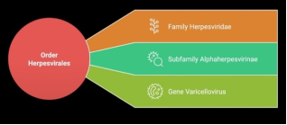
Figure 1. Taxonomy of Feline Herpesvirus 1 (FHV-1).
3. Virus Characteristics and Epidemiology
Alphaherpesvirus exhibits predictable biological behavior: it is notably species-specific, thus not a zoonosis, and is highly sensitive to common disinfectants, antiseptics, and detergents . It is inactivated within 3 hours at 37°C and within 5 minutes at 56°C; at 4°C, the virus remains infectious for approximately 5 months, and at 25°C for one month . The natural route of infection is direct contact with nasal, oral, and conjunctival secretions, with primary viral replication sites including the nasal septum mucosa, turbinates, nasopharynx, conjunctiva, and tonsils . The virus does not cause viremia, as it requires low-temperature epithelial tissues, such as those of the upper respiratory tract and conjunctiva, for replication . There is no evidence of transplacental transmission, although it has been demonstrated experimentally . In certain situations, such as catteries, indirect transmission can occur through contamination of food utensils, bedding, cleaning equipment, and personnel . Respiratory aerosol is not considered the primary mode of infection; however, macro-droplets expelled through sneezing can travel distances of 1-2 meters .
The highest viral expression occurs in kittens: herpetic lesions manifest in the postnatal period due to transmission of the virus from the queen to the kittens following viral reactivation triggered by the stress of parturition and lactation . Nevertheless, some queens may shed the virus near parturition without displaying any clinical signs of herpes . This serves as an effective mechanism for viral transmission to subsequent generations. The likelihood of kittens developing clinical symptoms depends on maternal derived antibodies (MDA); some kittens may experience subclinical infection and become virus carriers. The incubation period is approximately 2-6 days .
4. Virology and Pathogenesis
FHV-1 is predominantly epitheliotropic, with viral DNA rarely found in the corneal stroma. The virus causes necrosis of the respiratory, conjunctival, and corneal epithelia, with eosinophilic intranuclear inclusion bodies, frequent neutrophil presence, and fibrinous exudation . Severe cases involving the lower respiratory tract present with necrotizing to necrosuppurative bronchitis, bronchiolitis, and alveolitis with leukocyte infiltration. Epithelial cells exhibit a range from intact to degenerated, showing cytoplasmic swelling, apoptosis, or necrosis . Viral spread from the upper to lower respiratory tract results from viral dissemination through the airways .
The virus follows a dual cycle: a productive phase and a latency phase.
Productive “lytic” phase: The virus spreads through contiguous cells via endocytosis, releasing new virions near cell junctions. It has tropism for epithelial cells, which have extensive contacts with each other and neurons . According to Labib, herpes infection begins with glycoproteins B (gB), gC, gD, gH, and gL, crucial for host cell interaction. gB and gC bind to heparan sulfate proteoglycans on the host cell surface, initiating adherence . This interaction induces conformational changes facilitating gD binding to host receptors like nectin-1 in neurons and herpes virus entry mediators (HVEM) on epithelial cells. gH and gL form a multiglycoprotein complex enabling fusion of the viral envelope with the plasma membrane, allowing nucleocapsid entry. Viral protein 16 (VP16) triggers viral DNA replication via polymerase activation, leading to protein synthesis. Replication genes are categorized into pre-immediate early, immediate early, early, leaky late, and true late, promoting viral replication and capsid formation. Viral release occurs via exocytosis or cell lysis . Cytolysis results from the virus's intense replicative activity .
Latency phase: A symptom-free period without detectable virions in fresh tissues, where the virus can be reactivated under various stimuli, including organ culture . During latency, the virus persists as episomes primarily in sensory ganglia nuclei . Reactivation under stress involves anterograde axonal transport from nerves to peripheral sites, causing recrudescence and transmission . The main latency site is the trigeminal ganglia, though viral DNA is also found in the olfactory bulb, optic nerve, brain, and non-neural sites like ocular and respiratory tissues (turbinates, cornea, buccal mucosa, conjunctiva, salivary glands, and submandibular lymph nodes) .
Latency comprises three stages: implantation, maintenance, and reactivation.
(1) Implantation: This process begins during the acute phase of the disease, during viral replication in mucosal sites. Nerve endings retain the virus inside; the virus is transported through nerve axons by a movement called retrograde axonal transport. When the virus reaches the nerve ganglia, it infects nerve cells and other cells. This infection process lasts about a week.
(2) Maintenance: In nerve cells, the latency-associated transcript (LAT) gene is expressed, which is responsible for limiting nerve cell death by apoptosis and host cell lysis . LAT is a molecular marker of viral latency and responsible for inhibiting neuronal apoptosis and the reactivation factor . LAT is used to distinguish a persistent infection with low viral load from a latency phase. During the lytic phase, over 80 genes are expressed, while during the latency phase only the LAT gene is expressed . During the latency phase, no virions are detected . LAT regulates the ICP0 gene, essential for viral reactivation . Although viral DNA is found in the trigeminal ganglia and cornea , the LAT transcript has been found only in the ganglia, suggesting exclusive latency in these organs and not in the cornea . Of 39 corneas examined, none expressed the LAT gene, while in the trigeminal ganglia, 4 out of 16 examined expressed the LAT gene . An important role in inhibiting reactivation is attributed to CD8+ T lymphocytes .
(3) Reactivation/Recrudescence: The maintenance phase is transient; indeed, due to stressors, pharmacological or natural therapies, the virus can reactivate. Infectious virions follow the same nerve pathway to the periphery. The virus can then be easily detected by PCR on nasal, oral, or ocular samples. Neurons infected by latent virus, in which viral reactivation occurs, undergo lysis, which can lead to metaherpetic symptoms . Recrudescence refers to a reactivation of the virus with the presence of clinical symptoms . The persistence of the virus in cats was initially predicted based on epidemiological, circumstantial, and comparative studies, and later demonstrated in isolated subjects . About 82% of cats are carriers of the virus, with the possibility of recrudescence . Recrudescence occurs following chronic corticosteroid therapy, infectious diseases (FIV-FELV), some spontaneous situations, and stressful conditions such as moving to a new home, post-operative periods, childbirth and lactation , boarding, cat shows, mating, introducing another animal into the house, or changes in the surrounding environment . The administration of dexamethasone and prednisolone in hospitalized cats led to virus recrudescence in 69% of cases, with virus shedding starting from the fourth to the eleventh day. Moving to a new home led to virus recrudescence in 18% of examined cats; the shedding period varies between 4 and 10 days, continuing for 4-9 days. Spontaneous occasions (situations where the cat underwent no changes during the study period) occurred in 29% of cats. In 40% of cases, pregnant cats showed phases of recrudescence . Activation, duration, severity of the disease, and virus elimination depend on the state of the immune response. In particular, virus-shedding cats, after stress induced by a change of home, show differences compared to non-shedding cats:
a) They have a more severe acute infection than subjects that do not shed the virus.
b) They have higher stress levels (manifested by loss of appetite and nervous symptoms).
c) They have lower serum antibody levels .
5. Systemic Clinical Manifestation
(1) Acute manifestation:
a) Respiratory and systemic: Characterized by fever, anorexia, lethargy, sneezing, and serous nasal discharge that becomes mucopurulent within 5-7 days. Bronchial and bronchiolar involvement may cause cough and dyspnea . Rarely, the virus causes abortion, but generalized infections can be fatal, sometimes with neurological symptoms . Oral ulcers are more common in calicivirus infections. Although they have not been observed in germ-free infected cats, they are described in the literature and, when present, appear as ulcerative-necrotic lesions with focal hyperplasia and dysplasia of the buccal epithelium .
b) Facial dermatitis: Multifocal lesions, mainly on the nose and facial skin, appearing as vesicles, crusts, and ulcers . Oral or topical famciclovir showed good results in a case report (Figure 2).
(2) Chronic manifestation and recurrence:
a) Recurrence: Milder in adults and immunocompetent cats, including corneal ulcers, conjunctivitis, ocular discharge, sneezing, and nasal discharge .
b) Chronic rhinosinusitis: FHV damages turbinates, causing osteolysis in acute infection. PCR confirms viral presence in chronic rhinosinusitis . Famciclovir improved symptoms in two cats .
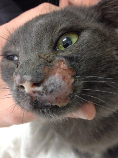
Figure 2. Facial dermatitis in a cat. (Courtesy of Dr. Necci F.).
6. Ocular Manifestations
FHV-1 is the most common feline ocular disease , with cytolytic, immunopathological, and metaherpetic mechanisms .
1. CYTOLYTIC MANIFESTATIONS (ACUTE AND RECURRENT SYMPTOMS)
a) Symblepharon: FHV-1 replicates in the epithelia of the conjunctiva and cornea and is responsible for necrotic-ulcerative inflammation and erosions in kittens. Contact between the corneal stroma and the conjunctival substantia propria is essential for the development of adhesions known as symblepharon (Figure 3). The presence of chemosis (not uncommon in some kittens with severe clinical manifestations) promotes contact between the surfaces . This condition is responsible for the reduction of the palpebral fissure (blepharophimosis), adhesions of the conjunctiva with the nictitating membrane, possible occlusion of the lacrimal ducts, difficulties in blinking, and keratoconjunctivitis sicca. In some cases, the entire cornea is covered by the conjunctiva, sometimes even pigmented (Figure 4, Figure 5). This predisposes to hypovision or possible blindness . Management of kittens with significant conjunctival hyperemia and chemosis should include the use of antivirals, antibiotic eye drops to reduce secondary infections, and functional eyelid movement to prevent the formation of adhesions .
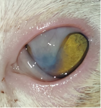
Figure 3. Symblepharon. (Courtesy of Dr. Naso S.).
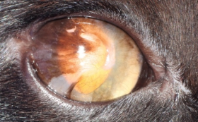
Figure 4. Lateral canthus symblepharon. Note the corneal neovascularization (chronic stromal keratitis). (Courtesy of Dr. Perez T.).
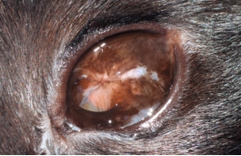
Figure 5. Pigmented symblepharon. (Courtesy of Dr. Perez T.).
b) Ulcerative keratitis: FHV-1 is considered the most common etiological agent causing corneal problems in cats . Excluding secondary causes of ulcers such as entropion, tear film abnormalities, eyelid neoplasms, foreign bodies, trauma, corneal infections, and rare cases secondary to ectopic cilia, distichiasis, or contact with caustic substances, all remaining ulcers can be attributed to herpetic causes, especially in kittens . In general, cats infected with herpesvirus mainly manifest punctate and dendritic ulcers. Dendritic ulcers are considered pathognomonic of herpetic infection, appearing as linear or branching epithelial lesions, sometimes with swollen edges (Figure 6). Dendritic ulcers are evaluated using vital stains like fluorescein and rose bengal (more sensitive) with the help of magnification systems such as a slit lamp . A dendritic ulcer can resolve or progress to geographic ulcers, which may also develop independently. In the first case, lesions resolve in approximately 2-3 weeks . In the second, coalescence of dendritic ulcers can form geographic ulcers due to infected epithelial desquamation and viral spread to the basement membrane . Geographic ulcers are extensive, superficial, with irregular borders resembling maps, often accompanied by edema and neovascularization (Figure 7) In some cases, the epithelium appears lifted, resembling canine SCCED erosions, but with different pathogenesis and clinical significance (Figure 8). In human herpes simplex, poor epithelial adherence is linked to neurotrophic damage and failure of regenerating epithelium to adhere to a damaged, slowly healing basement membrane . Both dendritic and geographic ulcers may occur during recrudescence, though accompanying conjunctival inflammation and ocular symptoms are milder . Corticosteroid use prolongs ulcer healing, with one study reporting a 29-day healing period without steroids compared to over 60 days with them . No veterinary literature addresses combined antiviral-corticosteroid use. There are no reference articles regarding whether epithelial removal is advisable. The epithelium detaches easily with the cat’s normal blinking or with the assistance of a cotton swab, although this maneuver can lead to sequestrum formation, as described in a clinical case by Stiles . Other methods, such as the Alger-brush, are not recommended due to the same risk of sequestrum formation. Bacterial infections can exacerbate ulcers into stromal ulcers, descemetoceles, or perforations, especially in kittens . Adult cats may show adherent leukomas with anterior synechiae from early-life perforations, not to be confused with persistent pupillary membranes (PPM) .
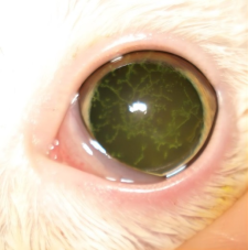
Figure 6. Dendritic corneal ulcer positive to fluorescein stain. (Courtesy of Dr. Tralhao P.).
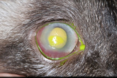
Figure 7. Chronic geographic ulcer. The ulcer is positive to fluorescein stain, with intense neovascularization and edema.
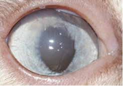
Figure 8. Geographic corneal ulcer without fluorescein stain. Note the central edema.
c) Conjunctivitis: Herpesvirus, along with chlamydia, mycoplasma, and calicivirus, is considered one of the etiological agents responsible for primary conjunctivitis . FHV is a cause of both acute and chronic conjunctivitis. An article highlights herpes positivity in 54% of cats tested with acute conjunctivitis . (Figure 9) The severity of conjunctivitis differs between a primary infection and a recurrence. In the primary infection, especially in kittens, conjunctivitis is characterized by intense hyperemia and chemosis, intense tearing, and purulent or serosanguineous discharge if conjunctival ulcers are present . Chemosis is not a constant finding and, when present, is never as evident as in other bacterial conjunctival infections caused by chlamydia . In some situations, viral conjunctivitis can become purulent following bacterial infections, both in acute and chronic forms . A study tested the presence of FHV-1 in 19% of subjects with chronic conjunctivitis through immunofluorescence . It is always recommended to inspect the nasal area during an ophthalmic examination in the presence of symptoms such as conjunctival hyperemia and tearing. It is also necessary to obtain a good medical history, perform a PCR to exclude other differential diagnoses, and confirm a respiratory infection to choose the correct therapy, either topical or systemic (Figure 10).
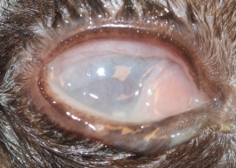
Figure 9. Acute herpes infection in a kitten. Conjunctivitis with purulent discharge.
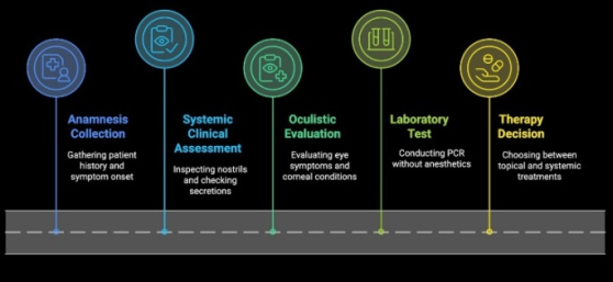
Figure 10. Synopsis of the management of infected subjects.
d) Ophthalmia neonatorum: It is a severe condition in kittens, in which a primary FHV-1 infection and a concomitant bacterial infection occur, potentially leading to serious corneal problems, up to ocular perforation with consequent phthisis . This condition occurs if the viral infection happens before the eyelids open, and degenerated inflammatory cells can accumulate in the palpebral space . Sometimes, in addition to swollen eyelids, pus can be seen oozing from the medial canthus. Therapy consists of early opening of the palpebral fissure, surface irrigation, and the use of antibiotic therapies .
2. IMMUNOPATHOLOGICAL ACTION AND HERPETIC-RELATED PATHOLOGIES WITH UNKNOWN CAUSES
a) Eosinophilic keratoconjunctivitis: A progressively evolving keratoconjunctivitis characterized by raised, white-pink, irregular perilimbal or peripheral lesions, staining with fluorescein, with edema and vascularization (Figure 11). Ulcers may develop before eosinophilic keratitis . Cytological examination reveals eosinophils, plasma cells, and lymphocytes . Possible mechanisms include: 1) IgE-mediated type I hypersensitivity with mast cell degranulation and chronic tissue damage from eosinophil degranulation; or 2) T-cell mediated type IV hypersensitivity with interleukin 5 production and local eosinophil response, leading to corneal damage . FHV-1’s role is unclear, though viral DNA was found in 76.3% and 54.5% of tested cats in two studies .
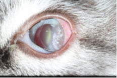
Figure 11. Eosinophilic keratitis. Note the white plaques. (Courtesy of Dr. Tralhao P.).
b) Corneal sequestrum: Necrotic corneal stroma appearing brown/black (Figure 12, Figure 13). The role of herpesvirus is unclear. Corneal sequestrum often accompanies chronic corneal ulcers, and herpesvirus involvement is evident . Viral DNA was found in 55.1% of cats with corneal sequestrum (overrepresented in Persian and Himalayan breeds), suggesting other contributing factors . Another study found no significant correlation between sequestrum and herpesvirus positivity via PCR . Surgical removal and antiviral therapy are recommended .
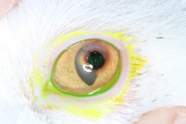
Figure 12. Corneal sequestrum in a cat. (Courtesy of Dr. Tralhao P.).
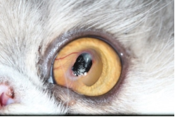
Figure 13. Corneal sequestrum. (Courtesy of Dr. Tralhao P.).
c) Uveitis: Widely considered a cause of chronic and recurrent uveitis in humans , its role is also suspected in chronic feline uveitis, given the biological similarity between the two viruses and the presence of lymphocytic infiltrates. In humans, uveitis is more frequent during episodes of recrudescence than in primary disease . The mechanism by which herpesvirus is responsible for uveitis is still unclear. Some suspect a direct cytolytic action of the virus, which reaches the inside of the eye through the sensory axons of the trigeminal nerve . Other authors suggest that herpetic uveitis might be an immunological reaction due to hypersensitivity to the viral agent. A study on herpes simplex in humans highlighted that intraocular inflammatory processes can result from infection with both inactivated and virulent viral agents . The only study in veterinary medicine found viral DNA, tested via PCR, in cases of chronic recurrent uveitis. Forty-four cats with idiopathic uveitis, 29 with uveitis attributed to Toxoplasma gondii, 13 herpes-seropositive cats without uveitis, and 9 seronegative cats without uveitis were tested. The virus was detected only in the aqueous humor of subjects with uveitis. Eleven out of twelve cats with chronic uveitis presented viral DNA in the aqueous humor. However, doubt remains as to whether FHV-1 is the causative agent of uveitis or a secondary infection to the disease .
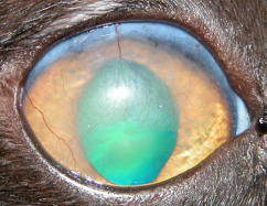
Figure 14. Chronic stromal keratitis in a cat. Note the intense fibrosis and neovascularization. (Courtesy of Dr. Vergara M.).
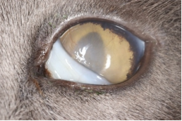
Figure 15. Chronic stromal keratitis.
d) Chronic stromal keratitis: In this clinical manifestation, more evident in cases of recurrent infections associated with recrudescence, the chronic stromal inflammatory process presents with fibrosis and neovascularization. In rare cases, edema due to endothelial damage may be observed (Figure 14, Figure 15). Chronic stromal keratitis is responsible for hypovision and rarely blindness . Studies on herpes simplex indicate that the insult is not directly induced by the virus but is an immunopathological consequence to stromal viral antigens . Supporting the immunopathological theory is evidence that a lower stromal reaction occurs in murine models where CD4+ T cells are depleted . Furthermore, in cases of chronic stromal ulcers, no virus can be detected; in fact, there is no active replicative phase, and the use of antivirals does not resolve the issue. The use of corticosteroids in this manifestation of the disease may be beneficial .
e) Keratoconjunctivitis sicca: Clinical signs include conjunctival hyperemia, ocular dryness, and ulcers . It is an occasional condition, seen as a consequence of acute or chronic manifestations. In acute forms, qualitative keratoconjunctivitis sicca is noted, with a drastic reduction of goblet cells, which even disappear in histological preparations by the seventh day post-infection . In the acute phase, however, excessive tearing is observed. In chronic or non-acute forms, quantitative keratoconjunctivitis sicca is known. The cause is attributed to obstruction of glandular ducts or secondary adenitis .
3. METAHERPETIC PATHOLOGIES (CHRONIC SYMPTOMS)
Metaherpetic diseases are related to functional damage affecting the cornea and ocular tissues, predisposing to chronic problems. They do not require antiviral therapy, as there is no active viral replication . Metaherpetic lesions are well described in human medicine but are poorly understood in veterinary medicine despite the frequency of the disease. A study investigated the lacrimal functional unit and corneal trigeminal nerve function in infected subjects . Cranial nerves play a crucial role in maintaining the lacrimal functional unit by stimulating blinking and lubrication, which are important factors for stimulating and promoting re-epithelialization. Herpesvirus is a neurotropic virus responsible for cytolysis of nerve cells as well. The results show a qualitative and quantitative decrease in the tear film, as well as a reduction in blinking frequency and corneal sensitivity. The latter is markedly reduced both with the use of the Cochet-Bonnet esthesiometer and in post-mortem studies of nerve fibers through confocal microscopy .
7. Immunity
(1) Passive immunity: Kittens with good passive immunity may remain asymptomatic but become latent carriers, spreading the virus . Colostrum provides passive immunity for 2-10 weeks, though antibody levels may sometimes be insufficient . Antibodies can persist beyond 10 weeks, but most kittens lose maternal antibodies (MDA) within 6 weeks . Some FHV-1 infected cats lack detectable serum antibodies but are protected from disease .
(2) Innate immunity: Viral glycopeptides activate innate immunity, triggering interferon production and activation .
(3) Active immunity: The infection does not provide solid immunity. Immunity protects against clinical symptoms but not against infection. Myeloid dendritic cells reach the lymph nodes, where they present the antigen to class 2 MHCs. This activates CD4+ helper cells, which undergo replication and differentiation into Th1 cells, producing interleukin 2 and IFN-γ, leading to the activation of CD8+ T cells . T cells therefore perform a dual action, both non-cytolytic and cytolytic, to mediate the clearance and destruction of infected cells, respectively. The non-cytolytic action is mediated by IFN-γ, which slows viral replication, while the cytolytic action induces cell apoptosis. Antibody production also occurs; however, since herpes cells require direct cell-to-cell contact for viral propagation, neutralizing antibodies often prove ineffective . Immunity has always been assessed through VNAs (virus neutralizing antibodies), although cellular immunity is certainly a more objective measure of immune status. In fact, cats with low antibody levels do not necessarily develop the disease . After a primary acute infection, VNA levels are low, sometimes imperceptible. After 6 months, protection may be only partial, and a recurrence may occur. After reactivation, VNAs increase and remain stable regardless of subsequent viral reactivation .
(4) Vaccines: Both live-attenuated and inactivated vaccines exist . They reduce symptoms but don’t prevent viral replication or latency . Vaccination around 9 weeks, with a second dose 3-4 weeks later, is recommended as MDA wane . Avoid contact with other cats pre-vaccination .
Vaccination guidelines:
a) Annual boosters for cats in multi-cat environments .
b) Boosters every 3 years for indoor-only cats .
c) Biannual boosters for breeding or boarding cats .
Vaccine reactions may include upper respiratory symptoms, ocular issues, and oral ulcers, typically within a week post-trivalent vaccination, likely due to calicivirus . Failures result from improper storage, administration, or immunocompromised status (systemic disease, immunodeficiency viruses, malnutrition, immunosuppressive therapy, stress, high viral loads in outbreaks) . High maternal antibody titers and premature vaccine schedules can also interfere . Live-attenuated vaccines may establish latency in trigeminal ganglia . Inactivated vaccines are preferred for pregnant or immunocompromised cats .
8. Diagnosis
a) Clinical: The presence of keratopathy associated with infectious inflammatory ocular manifestations is indicative of herpesvirus infection. Other respiratory agents do not cause corneal damage. Conversely, the presence of lingual or pharyngeal ulcers is typical of calicivirus infection and only rarely of herpesvirus . A thorough anamnesis and inspection of the nose and oral cavity can support the diagnosis.
b) Laboratory: The main laboratory diagnostic methods include PCR, virus isolation, and immunofluorescence. Cytology is rarely helpful in herpesvirus infections, as it is not always useful for visualizing intranuclear inclusion bodies and is considered supportive but not diagnostic. Neutralization tests and ELISA cannot differentiate between infection status (clinically normal, acute, or chronic) and vaccine immunity .
1) PCR
PCR allows amplification and identification of viral genomic fragments, enabling the detection of the virus with high sensitivity and specificity . Although it demonstrates the presence of the virus, it cannot assess active viral infection, as viral DNA can be found in the epithelia of asymptomatic cats . Various types of PCR have been studied, most targeting the viral thymidine kinase gene . Conjunctival scraping or peripheral corneal epithelium from the ulcer (not attached to the underlying stroma) can be used as samples for PCR analysis . Laboratories offer conventional PCR, nested PCR, and real-time PCR (qPCR). Nested PCR is used when increased sensitivity and/or specificity is required through two sequential amplification reactions, though conventional PCR results are often comparable .
Viral shedding and simultaneous qPCR evaluation allow differentiation of three phases:
(i) Phase one: Up to day 14 of infection, high viral titers and qPCR values are observed.
(ii) Phase two: Up to day 25, viral titers decline while PCR values remain high.
(iii) Phase three: After day 25, in the final phase of infection, viral titers are no longer present, and PCR values significantly decrease .
False positives: PCR detects viral DNA from active infection or degraded viral particles. Latent virus detection is unlikely as LAT presence in the cornea cannot be demonstrated . Viral DNA can be present in asymptomatic subjects or those with unrelated pathologies. Over 50% of asymptomatic subjects can shed the virus , meaning detection in a diseased subject could indicate coincidence, secondary infection, or true infection . Vaccination prevents clinical signs but not viral shedding during reactivation phases. False negatives: Despite PCR’s high sensitivity, false negatives may occur due to herpesvirus’s envelope sensitivity, DNA degradation during transport, sample quantity, collection site, or the use of ocular anesthetics , fluorescein, and rose bengal .
2) Virus isolation
Virus transport requires specific culture media. This method is less practical, requiring careful sample collection to avoid topically applied anesthetics that can degrade the virus . Once considered the "gold standard, " it has shown lower sensitivity than PCR in some studies .
3) Immunofluorescence
Recently less employed, it can be performed on conjunctival or corneal secretions collected using a cytobrush, Kimura spatula, or the blunt side of a scalpel . Fluorescein use prior to collection is discouraged due to interference with results . Sensitivity is limited by operator subjectivity .
9. Therapy
SUPPORTIVE THERAPY IN KITTENS
Supportive therapy includes fluid therapy and acid-base balance assessment. Many cats, due to the involvement of their sense of smell, do not eat, making it necessary to force-feed them with warm, palatable food. Appetite stimulants can be used, and if the cat still refuses to eat, the use of a feeding tube is necessary. Broad-spectrum antibiotic therapy is useful to prevent secondary infections. Nasal secretions must be cleaned, and the use of nasal decongestants, mucolytic agents (acetylcysteine), and nebulization with saline solutions can help liquefy the secretions . Oral antiviral medications (famciclovir) may also be helpful.
OCULAR THERAPY
1) ANTIBIOTICS: In cases of feline conjunctivitis and ulcerative keratitis, antibiotics are commonly used to reduce the risk of infections and symptomatic complications. Tetracyclines can be a good choice .
2) ANTIVIRALS: Before initiating antiviral therapy, it is important to consider the type and location of symptoms in the infected cat, whether solely ocular or also systemic. Antivirals are particularly effective in cases of acute infection but are less effective in cats with chronic stromal keratitis or corneal sequestrum . No antiviral has been specifically developed for cats; current options are derived from research on human herpes simplex virus. Some antivirals require host or viral modification to become active . Antivirals are virostatic and often require frequent administration. Their static action allows the host’s immune system to slow down the infection and mount an immune response. Antiviral agents can target any phase of viral replication, absorption, or release, though the most effective act on DNA replication .
a) Idoxuridine: A thymidine analog that competes for incorporation into viral DNA, rendering the virus unable to replicate. It is non-specific to the virus and affects any process requiring thymidine, including host processes; thus, it is used only topically . Idoxuridine 0.1% has proven useful when administered every 4-6 hours. In one study, idoxuridine was applied to 7 cats: two recovered, one improved, and four showed no improvement or worsened . The product is not commercially available but can be prepared as a compounded formulation.
b) Vidarabine: An adenosine analog that inhibits DNA synthesis by interfering with DNA polymerase. Like idoxuridine, it is non-selective for the virus and is preferably administered topically . Vidarabine requires administration 5-6 times daily and is not commercially available . In one study, vidarabine improved symptoms in 4 cats, but none achieved complete clinical resolution .
c) Trifluoridine: A thymidine nucleoside analog that likely reduces DNA synthesis by inhibiting thymidylate synthase. It penetrates the corneal stroma well but may cause irritation in treated cats. A 1% trifluoridine solution is used every 4-8 hours .
d) Cidofovir: A cytosine nucleoside analog that competes during DNA synthesis. Cidofovir 0.5% has shown efficacy in reducing clinical signs and viral replication (evaluated by real-time PCR). Although associated with nasolacrimal duct stenosis in humans, this has not been observed in cats . A recent study compared the efficacy of cidofovir, famciclovir, and ganciclovir in the treatment of herpetic corneal ulcers, identifying cidofovir as the most effective agent..
e) Acyclovir: An acyclic nucleoside analog that requires phosphorylation for activation, primarily by viral thymidine kinase. However, feline herpesvirus-1 thymidine kinase phosphorylates less efficiently than HSV-1, making acyclovir less effective in cats and potentially causing bone marrow suppression. Topical application is preferred. Acyclovir 0.5% ointment applied 5 times daily may help resolve pathology .
f) Ganciclovir: An acyclic nucleoside analog, also virostatic, requiring thymidine kinase-mediated phosphorylation . Available as a 0.15% gel formulation.
g) Famciclovir: A prodrug of penciclovir activated during gastrointestinal absorption. Dosages of 62.5 mg once daily for 10 days or 62.5 mg twice daily for 21-30 days yielded satisfactory clinical results in case studies . Another study reported excellent results with 90 mg/kg three times daily . Famciclovir has been used successfully in conjunctivitis, keratitis, sequestra, herpetic dermatitis, and rhinotracheitis . Penciclovir has a short plasma half-life and is excreted in urine. Although not nephrotoxic, dose adjustments are required in cats with renal insufficiency .
3) LYSINE: Highly debated for feline herpes with variable results. Lysine’s pharmacological activity appears linked to arginine depletion, an amino acid essential for viral replication, as lysine antagonizes arginine during replication. In vitro, viral replication significantly decreases only under conditions of low arginine. However, arginine is vital in cats, and its deficiency can cause hyperammonemia. High arginine levels in the presence of lysine may increase viral replication . A 2002 study found lysine well tolerated in cats, reducing conjunctivitis signs compared to placebo, though disease duration was similar between groups . Another study noted reduced viral shedding after relocation and concurrent use of 400 mg L-lysine . Conversely, some studies reported increased viral detection and more severe respiratory and ocular signs in lysine-treated cats compared to controls .
4) INTERFERONS: Cytokines involved in innate and cell-mediated immune responses, activating dendritic cells, enhancing antibody response, and T-cell/NK cell cytotoxicity . Interferons are classified into Type I (viral) and Type II (immune), with Type I further divided into α, β, γ, and ω groups . Viral presence triggers interferon production, inhibiting viral spread. In vitro studies show efficacy of human IFN-α and feline IFN-ω . Conjunctival MX protein, a marker of circulating interferon, was not detected with oral administration, though present in white blood cells . Pre-infection IFN-ω treatment showed no significant differences in symptoms or infection compared to controls . Similar results were found with human IFN-α2b and feline IFN-ω .
5) ARTIFICIAL TEARS: Due to prolonged Tear Film Break Up Time from goblet cell lysis and frequent corneal ulcers, artificial tears are essential. Preservative-free hyaluronic acid formulations improve Tear Film Break Up Time .
6) CORTICOSTEROIDS: Acute infections in cats receiving corticosteroids may develop stromal keratitis . Another study noted a case progressing to corneal sequestration . However, corticosteroids combined with antivirals improved one cat’s condition and resolved another’s pathology .
10. Management and Prevention
Pathology prevention involves vaccination and management.
1) Hospitalization and management of infected cats in veterinary facilities:
a) Gloves are mandatory during ocular examinations to prevent contamination.
b) If possible, create cat-only wards, separate from dogs and other animals that may cause stress .
c) Use disposable or sterile gloves when handling kittens and provide a heated environment.
d) Vaccinate kittens as early as possible, ideally at 4-5 weeks of age.
e) Separate kittens from mothers early (4-5 weeks) if necessary .
f) Handle infected cats carefully to avoid contamination from secretions. Infected cats should be isolated as sneezed particles can transmit the virus. Asymptomatic hospitalized cats can experience reactivation, requiring monitoring and isolation from other cats.
g) Minimize stress by enriching the environment with toys, familiar blankets, hiding spots, litter boxes, and palatable food .
h) Human interaction (play and grooming) increases S-IgA levels and reduces reactivation risk in latently infected cats .
i) Use Feliway® pheromone diffusers to reduce stress and respiratory symptoms .
j) Avoid small cages that restrict natural behaviors like stretching, walking, jumping, playing, and eating separately from the litter box .
k) Clean cages with common disinfectants and wait several days before introducing new cats .
2) Household Cats:
a) Vaccinate indoor cats with a known history of viral infections, even if they have no contact with other cats.
b) Minimize stress by creating cat-friendly environments (scratching posts) and using pheromone diffusers during changes.
c) Avoid relocating cats during owner absences; instead, arrange for daily care (feeding, litter changes) at home.
3) Management in Breeding Facilities, Colonies, and Boarding:
a) Unfamiliar handlers, disrupted feeding routines, breeding activities, unenhanced environments, adverse stimuli (noise, odors, inappropriate temperatures), and lack of hiding places can cause stress .
b) Quarantine new cats for about three weeks, separate from others, especially in non-disease-free facilities .
c) Perform PCR testing during quarantine .
d) Avoid breeding cats with herpes or a history of infected litters .
e) Isolate symptomatic cats to prevent transmission .
f) In colonies with endemic virus presence, manage through vaccination, stress reduction, and separating pregnant queens .
Abbreviations

FHV-1

Feline Herpes Virus

MDA

Maternal Derived Antibodies

VNA

Virus Neutralizing Antibodies

LAT

Latency-associated Transcript

Author Contributions
Roberto Lombardi is the sole author. The author read and approved the final manuscript.
Conflicts of Interest
The author declares no conflicts of interest.
References
[1] Maggs D. J., Clarke H. E. "Relative sensitivity of polymerase chain reaction assays used for detection of feline herpesvirus type 1 DNA in clinical samples and commercial vaccines." American Journal of Veterinary Research, 2005, 60, 1550-1555.
[2] Studdert M. J., Martin M. C. "Virus diseases of the respiratory tract of cats: 1. Isolation of feline rhinotracheitis virus." Australian Veterinary Journal, 1970, 46(3), 99-104.
[3] Crotti A., Barsotti G. "Congiuntiviti, cheratiti e cheratocongiuntiviti infettive nel gatto Parte 2: herpesvirus felino." Veterinaria, 2024, 38(2).
[4] Thomasy S. M., Maggs D. J. "A review of antiviral drugs and other compounds with activity against feline herpesvirus-1." Veterinary Ophthalmology, 2016, 19, 119-130.
[5] Maes R. "Felid herpesvirus type 1 infection in cats: a natural host model for alphaherpesvirus pathogenesis." ISRN Veterinary Science, 2012, Article ID 495830.
[6] Andrew S. E. "Ocular manifestations of feline herpesvirus." Journal of Feline Medicine and Surgery, 2001, 3, 9-16.
[7] Monne Rodriguez J. M., Leeming G., Kohler K., Kipar A. "Feline herpesvirus pneumonia: investigations into the pathogenesis." Veterinary Pathology, 2017, 54(6), 922-932.
[8] Rohowsky M. W., Griesemer R. A. "Experimental feline viral rhinotracheitis in the germfree cat." American Journal of Pathology, 1970, 58(2), 269-82.
[9] Gaskell R., Dawson S., Radford A., Thiry E. "Feline herpesvirus." Veterinary Research, 2007, 38, 337-354.
[10] Nasisse M. P., Guy J. S., Davidson M. G., Sussman W. A., Farley N. M. "Experimental ocular herpesvirus infection in the cat. Sites of virus replication, clinical features and effects of corticosteroid administration." Investigative Ophthalmology and Visual Science, 1989, 30(8), 1758-1768.
[11] Stiles J. "Feline herpesvirus." Veterinary Clinics of North America: Small Animal Practice, 2000, 30(5), 1001-1014.
[12] Maeda K., Horimoto T., Mikami T. "Properties and functions of feline herpesvirus type 1 glycoproteins." Journal of Veterinary Medical Science, 1998, 60(8), 881-888.
[13] Deng M., Liang H., Xu Y., Shi Q., Bao F., Mei C., Dai Z., Huang X. "Identification, genetic characterization, and pathogenicity of three feline herpesvirus type 1 isolates from domestic cats in China." Veterinary Sciences, 2024, 11, 285.
[14] Hamano M., Maeda K., Mizukoshi F., Une Y., Mochizuki M., Tohya Y., Akashi H., Kai K. "Experimental infection of recent field isolates of feline herpesvirus type 1." Journal of Veterinary Medical Science, 2003, 65(8), 939-943.
[15] Sheldon Tai S. H., Niikura M., Cheng H. H., Kruger J. M., Wise A. G., Maes R. K. "Complete genomic sequence and an infectious BAC clone of feline herpesvirus-1 (FHV-1)." Virology, 2010, 401, 215-227.
[16] Maggs D. J. "Update on pathogenesis, diagnosis and treatment of feline herpesvirus type 1." Clinical Techniques in Small Animal Practice, 2005, 20(2), 94-101.
[17] Thiry E., Addie D., Belak S., Boucraut-Baralon C., Egberink H., Frymus T., et al. "Feline herpesvirus infection. ABCD guidelines on prevention and management." Journal of Feline Medicine and Surgery, 2009, 11, 547-555.
[18] Hoover E. A. "Infectious disease of the dog and cat." In: Greene C. E. (Ed.), 4th Edition, Elsevier.
[19] Lombardi R. "Persistent pupillary membranes in dogs and cats." Veterinaria, 2012, 2, 97-104.
[20] Gaskell R. M., Povey R. C. "Experimental induction of feline viral rhinotracheitis virus re-excretion in FVR-recovered cats." The Veterinary Record, 1977, 12, 128-133.
[21] Gaskell R. M., Povey R. C., Transmission of feline viral rhinotracheitis. Vet Rec. 1982; 111: 359-362.
[22] Synowiec A., Dąbrowka A., Pachota M., Baouche M., Owczarek K., Niżański W., Pyrc K., Feline herpesvirus 1 (FHV-1) enters the cell by receptor-mediated endocytosis. J Virol. 2023; 97(8): e00117-23.
[23] Labin B. A., Chigbu D. I., Clinical management of herpes simplex virus keratitis. Diagnostics. 2022; 12(8): 2368.
[24] Dunn L. E., Birkenheuer C. H., Dufour R., Baines J. D., Immediate early proteins of herpes simplex virus transiently repress viral transcription before subsequent activation. J Virol. 2022; 96(22): e01222-22.
[25] Townsend W. M., Stiles J., Guptill-Yoran L., Krohne S. G., Development of a reverse transcriptase-polymerase chain reaction assay to detect feline herpesvirus-1 latency-associated transcripts in the trigeminal ganglia and corneas of cats that did not have clinical signs of ocular disease. Am J Vet Res. 2004; 65(3): 314-319.
[26] Wang Q., Zhou C., Johnson K., Colgrove R. C., Coen D. M., Knipe D. M., Herpesviral latency-associated transcript gene promotes assembly of heterochromatin on viral lytic-gene promoters in latent infection. Proc Natl Acad Sci U S A. 2005; 102(44): 16055-16059.
[27] Kennedy D. P., Clement C., Arceneaux R. L., Bhattacharjee P. S., Huq T. S., Hill J. M., Ocular HSV-1: is the cornea a reservoir for viral latency or a fast pit stop? Cornea. 2011; 30(3): 251-257.
[28] Jones C., Alphaherpesvirus latency: its role in disease and survival of the virus in nature. Adv Virus Res. 1998; 51: 81-133.
[29] Nasisse M. P., Glover T. L., Moore C. P., Detection of feline herpesvirus 1 DNA in corneas of cats with eosinophilic keratitis or corneal sequestrum. Am J Vet Res. 1998; 59(7): 856-858.
[30] Stiles J., McDermott B. S., Bigsby D., Willis M., Martin C., Roberts W., Use of nested polymerase chain reaction to identify feline herpesvirus in ocular tissue from clinically normal cats and cats with corneal sequestra or conjunctivitis. Am J Vet Res. 1997; 58(4): 338-342.
[31] Maggs D. J., Lappin M. R., Nasisse M. P., Detection of feline herpesvirus-specific antibodies and DNA in aqueous humor from cats with or without uveitis. Am J Vet Res. 1999; 60(8): 932-936.
[32] Liesegang T. J., Classification of herpes simplex virus keratitis and anterior uveitis. Cornea. 1999; 18(2): 127-143.
[33] Hargis A. M., Ginn P. E., Feline herpesvirus 1 associated facial and nasal dermatitis and stomatitis in domestic cats. Vet Clin North Am Small Anim Pract. 1999; 29(6): 1281-1290.
[34] Malik R., Lessels N. S., Webb S., Gotis-Graham P., Vitale C., Norris J. M., Power H., Treatment of feline herpesvirus-1 associated disease in cats with famciclovir and related drugs. J Feline Med Surg. 2009; 11(1): 40-48.
[35] Johnson L. R., Foley J. E., De Cock H. V., Clarke H. E., Maggs D. J., Assessment of infectious organisms associated with chronic rhinosinusitis in cats. J Am Vet Med Assoc. 2005; 227(4): 579-585.
[36] Hartley C., Aetiology of corneal ulcers. Assume FHV-1 unless proven otherwise. J Feline Med Surg. 2010; 12(1): 24-35.
[37] Gould D., Feline herpesvirus-1: ocular manifestations, diagnosis and treatment options. J Feline Med Surg. 2011; 13(5): 333-346.
[38] Stiles J., Treatment of cats with ocular disease attributable to herpesvirus infection: 17 cases (1983-1993). J Am Vet Med Assoc. 1995; 207(5): 599-603.
[39] Low H. C., Powell C. C., Veir J. K., Hawley J. R., Lappin M. R., "Prevalence of feline Herpesvirus 1, Chlamydophila felis, and mycoplasma spp DNA in conjunctival cells collected from cats with and without conjunctivitis." American Journal of Veterinary Research, 2007, vol. 68, no. 6, pp. 643-648.
[40] Hartmann A. D., Hawley J., Werckenthin C., Lappin M. R., Hartmann K., "Detection of bacterial and viral organisms from the conjunctiva of cats with conjunctivitis and upper respiratory tract disease." Journal of Feline Medicine and Surgery, 2010, vol. 12, pp. 775-780.
[41] Nasisse M. P., Guy J. S., Stevens J. B., English R. V., Davidson G., "Clinical and laboratory findings in chronic conjunctivitis in cats: 91 cases (1983-1991)." Journal of the American Veterinary Medical Association, 1993, vol. 203, no. 6, pp. 834-840.
[42] Dean E., Meunier V., "Feline eosinophilic keratoconjunctivitis: a retrospective study of 45 cases (56 eyes)." Journal of Feline Medicine and Surgery, 2013, vol. 15, no. 8, pp. 661-666.
[43] Prasse K. W., Winston S. M., "Cytology and histopathology of feline eosinophilic keratitis." Veterinary and Comparative Ophthalmology, 1996, vol. 6, no. 2, pp. 74-81.
[44] Stiles J., "Treatment of cats with ocular disease attributable to herpesvirus infection: 17 cases (1983-1993)." Journal of the American Veterinary Medical Association, 1995, vol. 207, no. 5, pp. 599-603.
[45] Chan N. S. W., Chee S. P., "Demystifying viral anterior uveitis: A review." Clinical and Experimental Ophthalmology, 2019, vol. 47, no. 3, pp. 320-333.
[46] Doymaz M. Z., Rouse B. T., "Immunopathology of herpes simplex virus infections." Current Topics in Microbiology and Immunology, 1992, vol. 179, pp. 121-136.
[47] Lim C. C., Reilly C. M., Thomasy S. M., Kass P. H., Maggs D. J., "Effects of feline herpesvirus type 1 on tear film break-up time, Schirmer tear test results, and conjunctival goblet cell density in experimentally infected cats." American Journal of Veterinary Research, 2009, vol. 70, no. 3, pp. 394-403.
[48] Sebbag L., Thomasy S. M., Leland A., Mukai M., Kim S., Maggs D. J., "Altered corneal innervation and ocular surface homeostasis in FHV-1-exposed cats: a preliminary study suggesting metaherpetic disease." Frontiers in Veterinary Science, 2021, vol. 7, article 1.
[49] Dawson E., Gaskell R. M., "Inconvenienti legati alla vaccinazione contro le infezioni respiratorie virali nel gatto." Veterinaria, 1995, anno 9, no. 1, pp. 105-109.
[50] Stiles J., Pogranichniy R., "Detection of virulent herpesvirus-1 in the corneas of clinically normal cats." Journal of Feline Medicine and Surgery, 2008, vol. 10, pp. 154-159.
[51] Green M. R., Sambrook J., "Nested polymerase chain reaction (PCR)." Cold Spring Harbor Protocols, 2019, vol. 2019, no. 1, pp. 175-178.
[52] Vögtlin A. C., Fraefel C., Albini S., Leutenegger C. M., Schraner E., Spiess B., Lutz H., Ackermann M., "Quantification of feline herpesvirus 1 DNA in ocular fluid samples of clinically diseased cats by real-time TaqMan PCR." Journal of Clinical Microbiology, 2002, vol. 40, no. 2, pp. 519-523.
[53] Goldschmidt P., Rostane H., Saint-Jean C., Batellier L., Alouch C., Zito E., Bourcier T., Laroche L., Chaumeil C., "Effects of topical anaesthetics and fluorescein on the real-time PCR used for the diagnosis of Herpesviruses and Acanthamoeba keratitis." British Journal of Ophthalmology, 2006, vol. 90, no. 11, pp. 1354-1356.
[54] Maggs D. J., "Antiviral therapy for feline herpesvirus infections." Veterinary Clinics of North America: Small Animal Practice, 2010, vol. 40, no. 6, pp. 1055-1062.
[55] Fontanelle J. P., Powell C. C., Veir J. K., Radecki S. V., Lappin M. R., "Effect of topical ophthalmic application of cidofovir on experimentally induced primary ocular feline herpesvirus-1 infection in cats" American Journal of Veterinary Research, 2008, 69, 289-293.
[56] Williams D. L., Robinson J. C., Lay E., Field H., "Efficacy of topical acyclovir for the treatment of feline herpetic keratitis: results of a prospective clinical trial and data from in vitro investigations" Veterinary Record, 2005, 157, 254-257.
[57] Thomasy S. M., Lim C. C., Reilly C. M., Kass P. H., Lappin M. R., Maggs D. J., "Evaluation of orally administered famciclovir in cats experimentally infected with feline herpesvirus type-1" American Journal of Veterinary Research, 2011, 72, 85-95.
[58] Maggs D. J., Collins B. K., Thorne J. G., Nasisse M. P., "Effects of L-lysine and L-arginine on in vitro replication of feline herpesvirus type-1" American Journal of Veterinary Research, 2000, 61, 1474-1478.
[59] Stiles J., Townsend W. M., Roger Q. R., Krohne S. G., "Effect of oral administration of L-lysine on conjunctivitis caused by feline herpesvirus in cats" American Journal of Veterinary Research, 2002, 63, 99-103.
[60] Maggs D. J., Nasisse M. P., Kass P. H., "Efficacy of oral supplementation with L-lysine in cats latently infected with feline herpesvirus" American Journal of Veterinary Research, 2003, 64, 37-42.
[61] Drazenovich T. L., Fascetti A. J., Westermeyer H. D., Sykes J. E., Bannasch M. J., Kass P. H., Hurley K. F., Maggs D. J., "Effects of dietary lysine supplementation on upper respiratory and ocular disease and detection of infectious organisms in cats within an animal shelter" American Journal of Veterinary Research, 2009, 70, 1391-1400.
[62] Rees T. M., Lubinski J. L., "Oral supplementation with L-lysine did not prevent upper respiratory infection in a shelter population of cats" Journal of Feline Medicine and Surgery, 2008, 10, 510-513.
[63] Maggs D. J., Sykes J. E., Clarke H. E., Yoo S. H., Kass P. H., Lappin M. R., Roger Q. R., Waldron M. K., Fascetti A. J., "Effects of dietary lysine supplementation in cats with enzootic upper respiratory disease" Journal of Feline Medicine and Surgery, 2007, 9, 97-108.
[64] Haid C., Kaps S., Gonczi E., Hassig M., Metzler A., Spiess B., Richter M., "Pretreatment with feline interferon omega and the course of subsequent infection with feline herpesvirus in cats" Veterinary Ophthalmology, 2007, 10(5), 278-284.
[65] Slack J., Stiles J., Leutenegger C. M., Moore G., Pogranichniy R. M., "Effects of topical ocular administration of high doses of human recombinant interferon alpha-2b and feline recombinant interferon omega on naturally occurring viral keratoconjunctivitis in cats" American Journal of Veterinary Research, 2013, 74, 281-289.
[66] Fulton R. W., Burge L. J., "Susceptibility of feline herpesvirus 1 and a feline calicivirus to feline interferon and recombinant human leukocyte interferons" Antimicrobial Agents and Chemotherapy, 1985, 28(5), 698-699.
[67] Bracklein T., Theise S., Metzler A., Spiess B. M., Richter M., "Activity of feline interferon-omega after ocular or oral administration in cats as indicated by Mx protein expression in conjunctival and white blood cells" American Journal of Veterinary Research, 2006, 67(6), 1025-1032.
[68] Nasisse M. P., English R. V., Tompkins M. B., Guy J. S., Sussman W. A., "Immunologic, histologic, and virologic features of herpesvirus-induced stromal keratitis in cats" American Journal of Veterinary Research, 1995, 56(1), 51-55.
[69] Tanaka A., Wagner D. C., Kass P. H., Hurley K. F., "Association among weight loss, stress, and upper respiratory tract infection in shelter cats." Journal of the American Veterinary Medical Association, 2012, vol. 240, no. 5, pp. 570-576.
[70] Worranan K., Jarudecha T., Rattanatumhi K., Ritchoo S., Muikaew R., Wangsud S., Sussadee M., "First study on stress evaluation and reduction in hospitalized cats after neutering surgery." Veterinary World, 2022, vol. 15, pp. 1-8.
[71] Gourkow N., Phillips C. J. C., "Effect of interaction with humans on behaviour, mucosal immunity and upper respiratory disease of shelter cats rated as contented on arrival." Preventive Veterinary Medicine, 2015, vol. 121, pp. 288-296.
[72] Contreras E. T., Hodgkins E., Tynes V., Beck A., Olea-Popelka F., Lappin M. R., "Effect of a pheromone on stress-associated reactivation of feline herpesvirus-1 in experimentally inoculated kittens." Journal of Veterinary Internal Medicine, 2018, vol. 32, pp. 406-417.
[73] M. A. Mironovich 1, A. Yoon, M. E Marino 1, N. E. Ineck, i Liu 1, R. T Carter 1, A. C Lewin 1 “Evaluation of compounded cidofovir, famciclovir, and ganciclovir for the treatment of feline herpesvirus ocular surface disease in shelter-housed cats” Vet Ophthalmol 2023 Apr: 26 Suppl 1: 143-153.
Cite This Article
  • APA Style

    Lombardi, R. (2025). Feline Herpesvirus: A Persistently Relevant Disease, from Infection to Clinical Management. American Journal of Laboratory Medicine, 10(4), 64-77. https://doi.org/10.11648/j.ajlm.20251004.11

    Copy | Download

    ACS Style

    Lombardi, R. Feline Herpesvirus: A Persistently Relevant Disease, from Infection to Clinical Management. Am. J. Lab. Med. 2025, 10(4), 64-77. doi: 10.11648/j.ajlm.20251004.11

    Copy | Download

    AMA Style

    Lombardi R. Feline Herpesvirus: A Persistently Relevant Disease, from Infection to Clinical Management. Am J Lab Med. 2025;10(4):64-77. doi: 10.11648/j.ajlm.20251004.11

    Copy | Download

  • @article{10.11648/j.ajlm.20251004.11,
      author = {Roberto Lombardi},
      title = {Feline Herpesvirus: A Persistently Relevant Disease, from Infection to Clinical Management
    },
      journal = {American Journal of Laboratory Medicine},
      volume = {10},
      number = {4},
      pages = {64-77},
      doi = {10.11648/j.ajlm.20251004.11},
      url = {https://doi.org/10.11648/j.ajlm.20251004.11},
      eprint = {https://article.sciencepublishinggroup.com/pdf/10.11648.j.ajlm.20251004.11},
      abstract = {Feline herpesvirus type 1 (FHV-1) is a widespread pathogen responsible for feline viral rhinotracheitis, a highly contagious disease affecting cats worldwide. This virus primarily targets the upper respiratory tract and ocular tissues, leading to clinical signs such as sneezing, nasal discharge, conjunctivitis, keratitis, and, in severe cases, corneal ulceration. FHV-1 is particularly problematic in young, immunocompromised, or stressed cats, and latent infections can result in recurrent clinical manifestations. The pathogenesis of FHV-1 involves initial viral replication in epithelial cells, followed by latency establishment in the trigeminal ganglia. Reactivation can occur due to stress, immunosuppression, or co-infections, leading to viral shedding and disease recurrence. Diagnosis is based on clinical presentation, supported by molecular techniques such as PCR, which detect viral DNA in ocular or respiratory secretions. Serological tests are less useful due to widespread exposure among the feline population. Management of FHV-1 includes supportive care, antiviral therapy, and immune modulation. Topical and systemic antivirals, such as famciclovir and cidofovir, have shown efficacy in reducing viral replication and alleviating clinical symptoms. Interferon therapy and other immunomodulatory treatments are under investigation to enhance antiviral responses. Lysine supplementation, previously recommended, has shown conflicting results in recent studies. Secondary bacterial infections are common and may require antibiotic therapy. Vaccination plays a crucial role in prevention, though it does not completely eliminate the risk of infection or reactivation. Stress management and maintaining optimal environmental conditions can help reduce recurrence. Ocular complications of FHV-1, including chronic conjunctivitis, symblepharon, corneal sequestrum, and stromal keratitis, pose significant challenges in feline ophthalmology. Despite advances in diagnosis and treatment, FHV-1 remains a significant concern in feline medicine. Understanding its pathogenesis, improving management strategies, and developing more effective preventive measures are essential for minimizing its impact on feline health.
    },
     year = {2025}
    }
    

    Copy | Download

  • TY  - JOUR
    T1  - Feline Herpesvirus: A Persistently Relevant Disease, from Infection to Clinical Management
    
    AU  - Roberto Lombardi
    Y1  - 2025/09/15
    PY  - 2025
    N1  - https://doi.org/10.11648/j.ajlm.20251004.11
    DO  - 10.11648/j.ajlm.20251004.11
    T2  - American Journal of Laboratory Medicine
    JF  - American Journal of Laboratory Medicine
    JO  - American Journal of Laboratory Medicine
    SP  - 64
    EP  - 77
    PB  - Science Publishing Group
    SN  - 2575-386X
    UR  - https://doi.org/10.11648/j.ajlm.20251004.11
    AB  - Feline herpesvirus type 1 (FHV-1) is a widespread pathogen responsible for feline viral rhinotracheitis, a highly contagious disease affecting cats worldwide. This virus primarily targets the upper respiratory tract and ocular tissues, leading to clinical signs such as sneezing, nasal discharge, conjunctivitis, keratitis, and, in severe cases, corneal ulceration. FHV-1 is particularly problematic in young, immunocompromised, or stressed cats, and latent infections can result in recurrent clinical manifestations. The pathogenesis of FHV-1 involves initial viral replication in epithelial cells, followed by latency establishment in the trigeminal ganglia. Reactivation can occur due to stress, immunosuppression, or co-infections, leading to viral shedding and disease recurrence. Diagnosis is based on clinical presentation, supported by molecular techniques such as PCR, which detect viral DNA in ocular or respiratory secretions. Serological tests are less useful due to widespread exposure among the feline population. Management of FHV-1 includes supportive care, antiviral therapy, and immune modulation. Topical and systemic antivirals, such as famciclovir and cidofovir, have shown efficacy in reducing viral replication and alleviating clinical symptoms. Interferon therapy and other immunomodulatory treatments are under investigation to enhance antiviral responses. Lysine supplementation, previously recommended, has shown conflicting results in recent studies. Secondary bacterial infections are common and may require antibiotic therapy. Vaccination plays a crucial role in prevention, though it does not completely eliminate the risk of infection or reactivation. Stress management and maintaining optimal environmental conditions can help reduce recurrence. Ocular complications of FHV-1, including chronic conjunctivitis, symblepharon, corneal sequestrum, and stromal keratitis, pose significant challenges in feline ophthalmology. Despite advances in diagnosis and treatment, FHV-1 remains a significant concern in feline medicine. Understanding its pathogenesis, improving management strategies, and developing more effective preventive measures are essential for minimizing its impact on feline health.
    
    VL  - 10
    IS  - 4
    ER  - 

    Copy | Download

Author Information